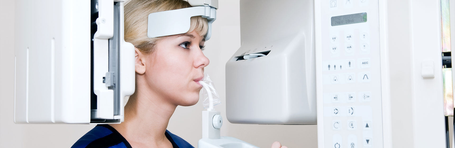

Orthophos XG 3D system is the latest technology that simplifies diagnostics and aids in treatment planning
Visitors to Longmont Dental quickly get to know the latest technologies that modern dentistry has to offer. Led by Dr. Kurt W. Knechtel, DMD, our team views technology as an essential ally. It supports our ability to provide a genuinely caring, highly relational, and personalized experience. The Orthophos® XG 3D complements skill at accurately diagnosing underlying conditions and developing effective treatment plans that result in the best outcomes, with the fewest side effects.
Brush up on the many benefits
This system represents one of the latest innovations debuted by Dentsply Sirona in the CBCT market. As a “cone beam computed tomography” scanner, the images produced by the Orthophos are captured at different angles. The machine features a specialized arm, which rotates around the patient a full 360 degrees. Cone beam imagery is known to be very clear and provides a “big picture” into both the oral cavity and the structures of the ear, nose, and throat. These diagnostic imaging technologies get their name from the fact that the x-ray beam is cone-shaped. These systems are also highly efficient, which makes them great for apprehensive patients or young and fidgety children.
Comprehensive scans can be rendered in a matter of seconds, and they can also be shared and viewed with the patient on a monitor. We feel this is a great way to build trust and relationships with patients because they are empowered with knowledge of the true nature and causes of their symptoms. This knowledge further informs their understanding of the best-recommended treatment options for them. They can make excellent and confident choices for their care that is also in their best interest.
Unique capabilities
The Orthophos® XG 3D system features two modes: standard and high-definition (HD) for endodontic treatments, such as root canal therapy. The standard renders a whopping 300 images in just one revolution, whereas the endo mode “bests” that – with 500 photos in one pass. Due to the built-in, highly-customizable features and capabilities, every aspect of the patient’s diagnostics and treatment planning can be altered. For instance, we can quickly and easily adjust the volume, dose, and quality associated with these images. The ability to pivot and personalize in such a way further supports the safety (by controlling the dose/radiation exposure) and the capture of very clear and detailed images of targeted areas efficiently and conveniently. Many other potentially complex cases and treatments, from orthodontics to implant placement, can further support this non-invasive diagnostic process.
Often, though, the technology has to be seen and experienced to understand and appreciate it. We encourage you to schedule a visit to our practice in Longmont. Please call (720) 815-4733.
We look forward to meeting you!


